|
|
 [Definitie:] "An ergogenic aid is any substance or phenomenon that enhances performance."
(Wilmore and Costill)
[Definitie:] "An ergogenic aid is any substance or phenomenon that enhances performance."
(Wilmore and Costill) 
|
|
|
||
|
||
|
0 2 - 0 1 - 2 0 0 6 Sudden anabolic steroid abuse-related death in athletes
Di Paolo M, Agozzino M, Toni C, Luciani AB, Molendini L, Scaglione M, Inzani F, Pasotti M, Buzzi F, Arbustini E.
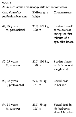
We describe the results of forensic autopsies and toxicological tests in four adult athletes (two professionals) who died suddenly out of the hospital.
The levels of testosterone were tested in urine samples using gas chromatographic mass spectrography (MS). The ratio between testosterone and its metabolite epitestosterone higher than six was considered as consistent with exogenous assumption of testosterone. Stanazolol was measured in urine samples using immunoenzymatic methods in ELISA. The presence of stanazolol was confirmed with HPLC/MS. The presence of cocaine, amphetamines, human chorionic gonadotropin (hCG), human growth hormone (hGH), insulin and other drugs was excluded. Alcoholic levels were within normal ranges. There were no prior medical history of illnesses, and of family history of cardiomyopathies, sudden death and coronary artery disease under the age of 50 years. The four athletes had regular evaluations for sport activity according to the Italian laws.
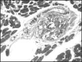
At autopsy, the four cases shared the following findings: absence of asymmetrical left ventricular hypertrophy, of coronary atherosclerosis causing significant luminal narrowing, pulmonary thromboembolism, coronary and endocavitary thrombi, and inflammatory infiltrates. Cardiac valves were normal. The histopathologic study showed myocardial damage characterised by myocyte hypertrophy, focal myocyte damage with myofibrillar loss, interstitial fibrosis, mostly at the subepicardial, and small vessel disease. Other organ/tissue findings were unremarkable. The cause of death was attributed as being cardiac due to the presence of myocardial changes similar to those seen in cardiomyopathies and small intramural artery disease and to the absence of other relevant pathological changes in other organs/tissues. In our four cases, the small arteriolar vessel disease was prominent. Small arteriolar vessels were thickened and showed intimal hyperplasia. These vascular lesions could play a role in causing chronic ischemic damage and arrhythmogenic potential. This small vessel disease could constitute a key point for future in vivo investigation with functional imaging tools.
|
|
|