 |
Twee capsules per dag zijn genoeg. Humaan onderzoek. (23.04.2024) |
Humaan onderzoek uit de koker van Sabinsa. Ondanks een welgemeende sponsor alert zeker interessant. (21.04.2024)
Afslanken met Immortality Herb zonder spierverlies
Zwaarlijvige mensen kunnen een lichaamsvet verliezen als ze elke dag 2 capsules met een extract van Immortality Herb of Gynostemma pentaphyllum innemen. (19.04.2024)
Intensieve beweging is een medicijn tegen kanker - maar teveel werkt averechts
Lichaamsbeweging is een superieure aanvullende behandeling bij kanker. Maar hoog-intensieve lichaamsbeweging is misschien wel iets wat wat je goed moet doseren. (17.04.2024)
Dit gebeurt er met je gezondheid als je vlees vervangt door vleesvervangers
Als je afgaat op wat langskomt in de media, zou je verwachten dat je gezonder wordt. Toch? (15.04.2024)
Wat gebeurt er als je wielrenners Pycnogenol, BCAAs, arginine en Robuvit geeft?
Gesponsorde en kleine studie. Toch interessant. (12.04.2024)
April
Dit doet een fikse dosis spirulina bij serieuze covid 10.04.2024
Langer leven door dierlijk eiwit 08.04.2024
Over Tongkat Ali en de seksualiteit van de mug 06.04.2024
Tekort aan omega-3-vetzuren fnuikt het veganistisch denkproces
04.04.2024
Oervoedsel vermindert kans borstkanker 02.04.2024
Maart
Spiermassa vasthouden tijdens een kort maar strikt dieet door suppletie met HMB 31.03.2024
Dagelijks gebruik van natto vermindert kans op gebroken bot 29.03.2024
Ook als je jong bent is trigonelline een natuurlijk anabolicum 25.03.2024
Trigonelline houdt je fit en sterk als de jaren klimmen 23.03.2024
Deeltijdvasten ontziet de botten 21.03.2024
Creatine heeft geen invloed op functioneren van nieren, zegt metastudie 19.03.2024
Klein handje noten versterkt de positieve effecten van training op de hersenen 17.03.2024
12 procent sterker, 15 procent fitter door urolithine A | Humaan onderzoek
15.03.2024
Cordyceps sinensis voor hardlopers 13.03.2024
Vitamine K2-MK7 beschermt botten tegen afbraak 11.03.2024
Hoe ouder je bent, hoe belangrijker is suppletie met vitamine D 09.03.2024
Na een zware training herstellen sporters die weinig zitten sneller 07.03.2024
Meer spieren, minder vet door knoflook 05.03.2024
Zwarte thee remt kanker van prostaat sterker dan groene thee 03.03.2024
Over enokitake, testosteron en adenosine 01.03.2024
Februari
Trainen na eiwitshake verbrandt net zoveel vet als trainen op nuchtere maag 26.02.2024
Passiflora incarnata maakt het doorbreken van verslaving aan opiaten dragelijker 24.02.2024
Waarom zestigers met een doel in het leven gezonder zijn 22.02.2024
Reishi | Een spaak in de wielen van agressieve borstkanker 20.02.2024
Meer vitamine B3 geeft 40-plusser meer spieren en minder lichaamsvet 18.02.2024
Als angstremmer werkt Passiflora incarnata net zo goed als oxazepam 16.02.2024
Supplement met vitamine D3 reduceert pijnklachten door fibromyalgie 14.02.2024
Slapeloze nachten? Supplement met Passiflora incarnata maakt ze korter 12.02.2024
Hoe koffiedrinkers zichzelf beschermen tegen Covid-19 10.02.2024
Chaga remt longkanker 08.02.2024
Supplement met berberine maakt bijna-diabeet gezonder en slanker 06.02.202 Shot met kurkuma en vitamines reduceert aantal ziekdagen topsporters 04.02.2024
Vitamine D als pijnstiller 02.02.2024
Januari
Visolie onttobt, ontzorgt en vermindert angst 31.01.2024
Echinacea beschermt tegen virussen in het algemeen, niet tegen SARS-CoV-2 in het bijzonder 29.01.2024
Meer en beter werk afleveren? Slaap en beweeg meer 27.01.2024
De onvermoede antivirale werking van Sint Janskruid 25.01.2024
Metastudie bevestigt bescherming quercetine tegen Covid-19 23.01.2024
Suppletie met N-acetyl-D-glucosamine brengt NK-cellen in stelling tegen griepvirussen 21.01.2024
Co-suppletie met gember + echinacea versterkt reguliere behandeling tegen Covid-19 19.01.2024
Goede voeding beschermt 65-plusser tegen Covid-19 15.01.2024
Zo sterk is de antidiabete werking van fenegriek 12.01.2024
Zoetstof erythritol helpt diabeten aan meer testosteron | Dierstudie 10.01.2024
Dit is het effect van ordinaire zwarte thee op je cholesterol 08.01.2024
Een zinvol leven? Minder kans op dementie 06.01.2024
Astaxanthine maakt mannen vruchtbaarder 04.01.2024
Astaxanthine voor wielrenners 02.01.2024
December
Versterken vitamine C en arginine elkaars antivirale werking? 29.12.2023
Dit doet een dagelijkse dosis van vier gram arginine en een gram vitamine C met long Covid 27.12.2023
Yoghurteters zijn slanker en hebben een lagere bloeddruk 25.12.2023
Waarom er in biologisch geteelde spinazie meer anabole ecdysteroïden zitten 23.12.2023
Versneld herstel en minder complicaties na hartoperatie door extra vitamine C 21.12.2023
Beroerte overleefd? Zorg voor voldoende magnesium 19.12.2023
Dit voedingspatroon verbetert misschien de vruchtbaarheid van vrouwen 17.12.2023
Je huid gezonder maken met collageen? Zo weinig heb je nodig... 15.12.2023
Bèta-alanine tegen opvliegers 13.12.2023
Sneller reageren met minder stress door Lion's mane 11.12.2023
Dit gebeurt er met je IGF-1-spiegel als je elke dag 2 eieren eet 09.12.2023
Tongkat Ali helpt hypogonadale mannen, concludeert metastudie 07.12.2023
Supplement met Pycnogenol en L-arginine verbetert seksueel functioneren van mannen 05.12.2023
Ashwagandha helpt bij vermoeidheid, niet bij stress 03.12.2023
Na afslankdieet is dikke man mannelijker | Metastudie 01.12.2023
November
Bij behandeling depressie overtreft Sint Janskruid werking SSRIs 29.11.2023
Supplement met vitamine E ontlast de vervette lever 27.11.2023
Fors minder pijn na operatie door supplement met vitamine C 25.11.2023
50 milliliter wortelsap erin, een kilo lichaamsvet eraf 23.11.2023
Leverwaarden verhoogd? Misschien helpt curcumine 21.11.2023
Hoe meer kilometers je rent, hoe groter je behoefte aan omega-3-vetzuren 19.11.2023
Creatine versnelt herstel long Covid 17.11.2023
Bij artritis houden champignons het immuunsysteem in toom 15.11.2023
Je lichaam gaat langer mee als je zorgt voor voldoende zink 13.11.2023
Het tweevoudig-remmende effect van rutine op het coronavirus 11.11.2023
Volledige baan + stress = sneller verouderen 08.11.2023
Wil je 12 jaar langer leven? Ga hardlopen (of joggen) 06.11.2023
Rutine | Misschien de meest effectieve remmer van Covid-19 in propolis 04.11.2023
Geveld door Covid-19? Braziliaanse propolis verbetert je immuunsysteem 02.11.2023
Oktober
Tai-Chi vertraagt verloop van de ziekte van Parkinson 29.10.2023
Aromatherapie tijdens de slaap voor een beter geheugen 27.10.2023
Sociale activiteit beschermt tegen dementie 25.10.2023
Wortelsap legt vrije radicalen aan de leiband 23.10.2023
Een komkommer per dag voor een lagere bloeddruk 21.10.2023
De proseksuele werking van saffraan 19.10.2023
Dit is het effect van suppletie met creatine op lichaamsvet 17.10.2023
Door het remmen van AGEs vertraagt carnitine misschien veroudering 15.10.2023
Volgens metastudie van 13 metastudies verbetert carnitine cholesterol 13.10.2023
De mogelijke antidepressieve werking van een supplement met vitamine D 11.10.2023
Minder kans op nasleep Covid-19 door visolie 09.10.2023
Zo zet gember je immuunsysteem een tandje hoger 07.10.2023
Bot gebroken, extra zink? 05.10.2023
Inpassen van walnoten in dieet verlaagt bloeddruk 03.10.2023
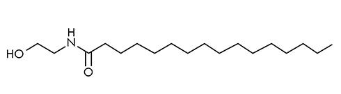
Je slaapt eerder in en je wordt 's morgens beter wakker door PEA 01.10.2023
September
Een dipeptide voor een langer leven met minder ziekte? | Thymogen 29.09.2023
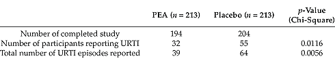
PEA tegen chronische pijn | Metastudie 27.09.2023
Metastudie | Meer Q10, minder vermoeid 25.09.2023
Zo goed werkt Ginkgo biloba tegen angst 23.09.2023
Dertig gram oesterzwam per dag verlaagt bloeddruk, vergroot gevoeligheid insuline 21.09.2023
Combinatie van dierlijke en plantaardige producten houdt spieren 65-plussers optimaal gezond 19.09.2023
Na de overgang maakt een dagelijkse theelepel olie van pompoenzaad vrouwen gezonder 17.09.2023
Thymogen versnelt genezing wonden bij diabetes | Laat het ook sporters beter presteren? 15.09.2023
Een beter geheugen door creatine 13.09.2023
Citrusfenol hesperidine vermindert spierafbraak door veroudering 11.09.2023
Vis houdt je fit na je 65ste 09.09.2023
Noten beschermen je gezondheid als de jaren klimmen 07.09.2023
Pijnlijke knie? Calciumfructoboraat helpt al binnen enkele dagen 05.09.2023
41 procent meer duurvermogen na korte monsterkuur ergothioneïne 03.09.2023
Wortels, sla, kool | Deze groenten beschermen je lever 01.09.2023
Augustus
Hoge inname vitamine E houdt hersenen 60-plussers fit 28.08.2023
Elke dag een paar noten, minder kans op depressie 25.08.2023
Trouw elke dag een supplement met vitamine D innemen halveert kans op melanoom 23.08.2023
Ook sporters die dagelijks koffie drinken reageren op suppletie met cafeïne 21.08.2023
210 of 420 milligram - welke dosis cafeïne werkt beter? 19.08.2023
Verhoging EPO-spiegel door ketonen vergelijkbaar met training in de bergen 17.08.2023
Whey maakt diabeten gezonder 15.08.2023
Vitamine C behoedt knie voor slijtage 13.08.2023
Al bij een verrassend geringe inname hebben Aloë vera-sterolen een positief effect op de huid 11.08.2023)
Eet kiwi's en je verkoudheid is sneller over 09.08.2023
Lichaam gaat beter om met glucose door kombucha 05.08.2023
Hogere consumptie van vlees, langere levensduur 03.08.2023
Glyoxylzuur, een metaboliet van glycine die spieren laat groeien 01.08.2023
Juli
Een warm bad voor de maaltijd verhoogt afgifte groeihormoon 30.07.2023
Na hartinfarct houdt megadosis choline hartpatiënt in leven 28.07.2023
Versnelt Spilanthes acmella de spiergroei van bodybuilders? Maar hoe dan? 26.07.2023
Pycnogenol maakt haardos voller 24.07.2023
Supplementen met visolie beschermen tegen coronaire hartziekten 21.07.2023
Chlorella is een uitstekende bron van carotenoïden 19.07.2023
Acetylcarnitine verdrijft vermoeidheid 17.07.2023
Komkommers tegen kanker? 15.07.2023
Een bèta-2-agonist die (misschien) lichaamsvet laat verdwijnen en spiermassa helpt opbouwen | Tripeptide Arg-His-Trp 13.07.2023
Alpha-Lipoic Acid reduceert pijn zonder oorzaak 11.07.2023
Spirulina vergroot misschien fertiliteit van mannen 09.07.2023
Immuunsysteem beter bestand tegen infectie door suppletie met creatine 07.07.2023
Chondroïtine houdt hartpatiënt in leven | De Morrisonstudie 05.07.2023
Chondroïtine reduceert kans op hartinfarct met een slordige veertig procent 03.07.2023
Gember imiteert werking insuline | Is gemberthee een supplement voor sporters? 01.07.2023